EVIS X1 Video System Center
CV-1500

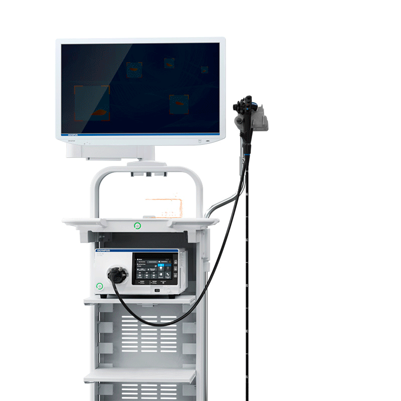
EVIS X1 Video System Center
The EVIS X1 CV-1500 Video System Center combines video processor and LED light source in one box, resulting in a more compact and lightweight design. Additionally, cross compatibility between EVIS EXERA III and EVIS LUCERA ELITE lead to a vastly extended portfolio of compatible endoscopes. The CV-1500 features multiple special observation modes that support physicians in every step during endoscopic procedures.

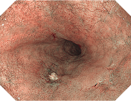
Texture and Color Enhancement Imaging (TXI)
Early detection is critical for cancer prevention and decreasing mortality.1 However, precursor lesions are often tiny and easy to overlook.
TXI technology aims to enhance the visibility of potentially suspicious tissue, which includes inflammations, flat or depressed lesions, using a white-light imaging effect that improves the color, structure and brightness.
By supporting better visibility of potential lesions, TXI aims to contribute to higher detection rates.

White Light
TXI

Texture and Color Enhancement Imaging
The incoming image is split and the texture and brightness are enhanced before the separate images are merged back together. Additional color enhancements are made to more clearly define subtle tissue differences.

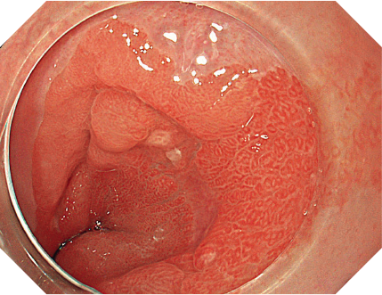
Narrow Band Imaging (NBI)
Accurate optical diagnosis is important when assessing lesions to determine potential histology, confirm the lateral extent, and thereby guide therapy decisions and suitable patient surveillance intervals.
NBI is a powerful and proven optical technology that allows for a reliable optical diagnosis of major indications in the gastrointestinal tract.1-8
Efficient lesion management strategies that are empowered by NBI include:
White Light
NBI

Narrow Band Imaging
Utilizing specific blue and green wavelengths absorbed by hemoglobin, NBI creates a strong contrast between vessels and surrounding mucosa.9 This facilitates the visibility of highly vascularized areas, blood vessel patterns and surface structures that are predictive for distinct histopathologies.10-13

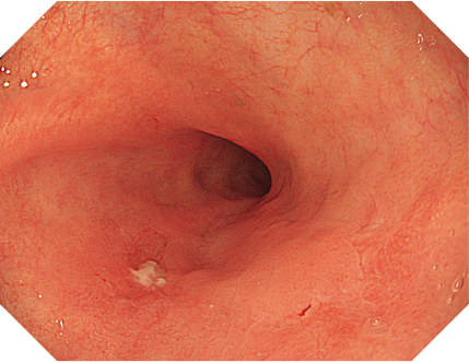
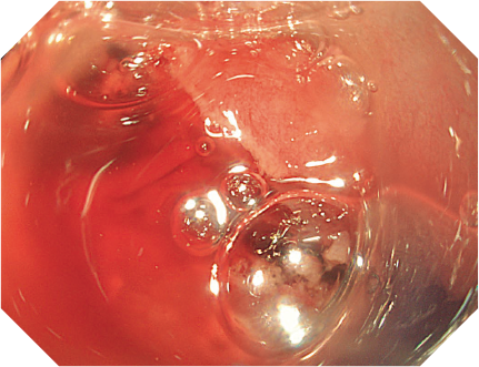
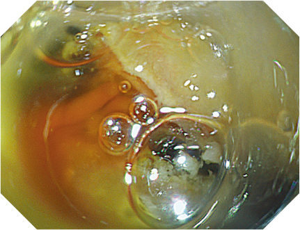
Red Dichromatic Imaging (RDI)
Gastrointestinal bleeding is a serious challenge, involving considerable mortality of 5-15% and high management costs.
1,2 Consequently, prevention of complications is crucial.
RDI is designed to enhance the visibility of deep blood vessels and bleeding sources.
Developed to facilitate fast, accurate identification and localization of bleeding sites, RDI may help perform speedier, more reliable hemostasis with the aim of reducing stress on physicians.
White Light
RDI

Red Dichromatic Imaging
RDI works by employing specific green, amber and red wavelengths. The latter two penetrate deep into the mucosa, enabling the visualization of deep blood vessels. In case of acute bleeding, RDI increases the contrast between highly concentrated and diluted blood, thereby clearly visualizing the bleeding point.
One dedicated platform: EVIS X1 merges the two worlds of EVIS EXERA III and EVIS LUCERA ELITE into one —resulting in a vastly extended portfolio of compatible endoscopes.
In the field of AI, we recognize the power to elevate endoscopic imaging to uncharted levels. The next upcoming feature of the EVIS X1 system will be computer-aided diagnosis (CAD) using artificial intelligence.

EVIS X1 provides diagnostic and therapeutic innovation alongside proven technologies to improve endoscopic procedures and endoscope handling.
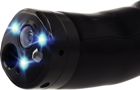
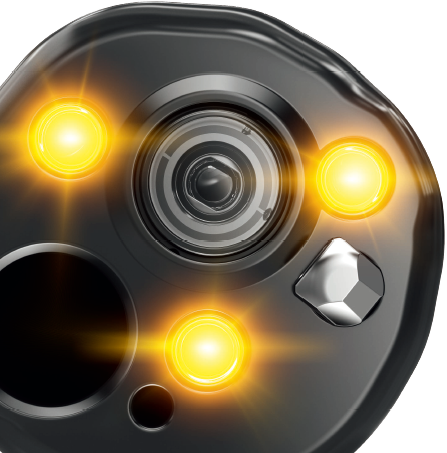
The EVIS X1 Video System Center contains five LEDs that are combined to produce different observation modes. It includes an Olympus tailored amber LED enabling the visualization capabilities of the RDI mode. LEDs have a longer life span than other light sources while consuming less energy.
BAI-MAC (Brightness Adjustment Imaging with MAintenance of Contrast) is a new image processing technique to improve the brightness levels in dark areas of the endoscopic image, whilst maintaining the brightness of lighter areas to increase the distance view.
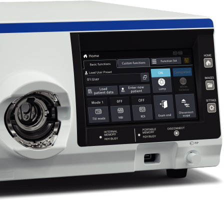
The EVIS X1 Video System Center is operated from a touch panel on the front of the unit, allowing the user to initiate all procedures and settings and control the image data from one device.
Pre-freeze analyzes previous images to obtain a clear visual record of the procedure in the shortest possible time (this is an updated algorithm compared to the EVIS EXERA III / EVIS LUCERA ELITE systems).
| Power Supply | Rated voltage | 100-240 V AC; Within ±10% |
|---|---|---|
| Frequency | 50/60 Hz; within ±3 Hz | |
| Rated input | 600 VA | |
| Size | Dimensions (W x H x D) | 370 x 198 x 488 mm; 398 x 218 x 580 mm (maximum) |
| Weight | 19.4 kg | |
| Classification (Medical Electrical Equipment) | Type of protection against electric shock | Class I |
| Degree of protection against electric shock of applied part | Depend on applied part. (The degree of protection against electric shock of this product is BF type if the mounting part to be connected to this product is BF type. However CF type is not subject to combination in this product.) | |
| Degree or protection against explosion | The video system center should be kept away from flammable gases. | |
| Observation | Analog signal output | VBS composite |
| Digital signal output | 12G-SDI (SMPTE ST 2082), 3G-SDI (SMPTE424M), HD-SDI (SMPTE292M), SD-SDI (SMPTE259M) | |
| User settings | The function settings for up to 20 users can be stored. | |
| Color tone adjustment | Adjust the color tone of each endoscopic image for White light observation mode, NBI observation mode, and RDI observation mode. · Red adjustment : ±8 steps · Blue adjustment : ±8 steps · Chroma adjustment : ±8 steps |
|
| Automatic gain control (AGC) | The image can be electronically amplified when the light is inadequate due to the distal end of the endoscope being too far from the object. | |
| Contrast | · H (High) : Darkens the dark part and brightens the bright part. · L (Low) : Brightens the dark part and darkens the bright part. | |
| BAI-MAC | Brightness adjustment with maintenance of contrast | |
| Iris | The iris modes can be switched. · Auto : The brightness is adjusted based on the brightest part of the central part and the average brightness of the periphery part. · Peak : The brightness is adjusted based on the brightest part of the endoscopic image. · Average : The brightness is adjusted based on the average brightness of the endoscopic image. | |
| Image enhancement settings | Fine patterns or edges in the endoscopic images can be enhanced electrically to increase the image sharpnes. · Enhancement type A : Emphasizes the pattern and contour of the endoscopic image. · Enhancement type B : Emphasizes the finer parts than structure emphasis type A. |
|
| Switching the enhancement modes | The enhancement level can be selected from 3 levels (OFF, 1, 2, and 3) | |
| Image size selection | The size of the endoscopic image can be selected from 2 modes. (Except SDTV) | |
| Electric zoom | Switch between mode 1, mode 2, and mode 3. | |
| PIP/POP | Switch between PIP and POP. | |
| Aspect ratio | Switch between 16:9 and 4:3. (Except SDTV) | |
| Freeze | Freeze the endoscopic image. | |
| Pre-freeze | The image with the least blur is selected from the images captured in the set time period before freeze operation and displayed. | |
| Optical-digital observation | The optical-digital observation can be performed. The endoscope compatible with the optical-digital observation is required. · NBI observation : This observation mode uses the narrow band light. · RDI observation : This observation mode uses the red dichromatic lights. · AFI observation : This observation mode uses the blue light. · TXI observation : This observation mode enhances color, texture and brightness. |
|
| Beginning and ending examination | Beginning and ending examination timing can be set interlock with the particular operation. | |
| Custom switch | Assign specific functions to the following buttons. · Remote switches (Up to 5) · Foot switches (Up to 2) · Keyboard custom key (Up to 4) · Touch panel custom button of basic functions screen (Up to 3) · Touch panel custom button of custom functions screen (Up to 10) | |
| MyCV mode | Switch setting values of multiple functions at once. | |
| Documentation | Remote control | The following peripheral device can be controlled (specified models only). · Portable memory · Video recorder · Color video printer · Image filing system · Server |
| Patient information | The following data can be displayed on the monitor. · Patient ID · Patient name · Gender · Age · Date of birth · Comment | |
| Displaying the record state | The recording state of the following peripheral device can be displayed on the monitor. · Portable memory : Remaining capacity · Video recorder : Number of shots / Recording status · Color video printer : Number of shots · Image filing system : Number of shots |
|
| Displaying the image information | The following data can be displayed on the monitor. · Image enhancement · Electric zoom ratio · Color mode · Focus · Observation mode | |
| Advanced registration of patient information | Up to 50 patient information can be registered. · Patient ID · Patient name · Gender · Age · Date of birth | |
| Recording format | Standard image quality: TIFF; Low image quality: JPEG | |
| Memory Backup | Memorization of user settings | The settings are held in memory even after the video system center is turned OFF. |
| White balance | The white balance that is once set is held in memory (only when using the compatible endoscope). |
Contents end

 A Unified Platform with 5 LED Spectrum Technology
 BEYOND VISION
 Revolutionary Integrated Bipolar and Ultrasonic Surgical Instrument with Enhanced Safety
 GET CLOSER